











Capnography is a core component of modern veterinary anesthesia monitoring. By measuring carbon dioxide in respiratory gases and displaying both a numeric value and a waveform, capnography gives the anesthesia team continuous information about ventilation, airway integrity, breathing-circuit performance, and pulmonary perfusion.
For small-animal patients, this information matters. Cats, toy-breed dogs, pediatric patients, and other small patients can deteriorate quickly, making continuous ventilation monitoring especially important. Changes in ventilation may appear on an EtCO₂ monitor before oxygen saturation or other parameters change. Used correctly, veterinary capnography helps the team recognize developing problems, investigate their possible causes, and escalate concerns promptly.
This guide explains the fundamentals of capnography for small animals and provides a practical approach to setup, interpretation, and troubleshooting.
> Safety disclaimer: This article is for educational purposes only. It does not replace the attending veterinarian’s clinical judgment, hospital protocols, current professional guidance, or the instructions provided by the equipment manufacturer.
What Do Capnography and EtCO₂ Measure?
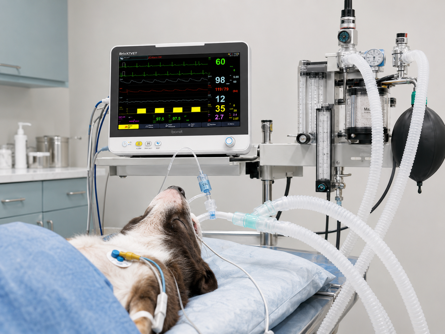
Capnography is the continuous measurement and graphic display of carbon dioxide in respiratory gases. The display typically includes:
- A numeric end-tidal carbon dioxide value, or EtCO₂
- A respiratory rate
- A continuous capnogram, or carbon dioxide waveform
- In some systems, inspired CO₂ and anesthetic agent measurements
EtCO₂ is measured near the end of exhalation, when the sampled gas most closely represents gas leaving the alveoli. In a well-perfused patient, EtCO₂ can provide a useful estimate of alveolar carbon dioxide and can help assess the relationship between ventilation and pulmonary perfusion. EtCO₂ does not directly measure arterial carbon dioxide. In many patients, PaCO₂ is higher than EtCO₂, and the difference may widen with pulmonary disease, impaired perfusion, ventilation-perfusion mismatch, or increased dead space. [1][2]
A single EtCO₂ value is useful, but the waveform and trend are equally important. A rising number with a consistent waveform may suggest hypoventilation. A sudden loss of waveform may indicate apnea, airway disconnection, accidental extubation, or equipment failure. A baseline that does not return toward zero may indicate rebreathing.
The 2025 ACVAA Small Animal Anesthesia and Sedation Monitoring Guidelines include continuous time-based capnography with waveform display as an important component of ventilation monitoring for anesthetized small-animal patients.[2] It should be used with hands-on patient assessment and other monitoring parameters.
Why Veterinary Capnography Matters
Capnography supports assessment of several connected systems:
Ventilation
EtCO₂ reflects how effectively the patient is eliminating carbon dioxide. Reduced respiratory rate, shallow breathing, excessive anesthetic depth, increased dead space, or inadequate ventilator settings can cause EtCO₂ to rise.
Airway integrity
A persistent waveform confirms that exhaled gas is reaching the sensor. An absent or suddenly changed waveform should prompt immediate assessment of endotracheal tube position, patency, and connection to the breathing circuit.
Breathing-circuit function
Capnography can help identify leaks, disconnections, rebreathing, exhausted absorbent, malfunctioning valves, and inadequate fresh-gas flow.
Pulmonary perfusion
When ventilation remains relatively stable, a sudden fall in EtCO₂ can reflect reduced pulmonary blood flow or cardiac output. A marked decrease may occur with severe hypotension, poor perfusion, or cardiopulmonary arrest.
Early deterioration
Pulse oximetry primarily evaluates oxygenation and may remain acceptable while ventilation is worsening, particularly when supplemental oxygen is being delivered. Capnography provides a direct, breath-by-breath view of ventilation and may reveal a developing problem earlier.
Understanding a Normal Capnogram
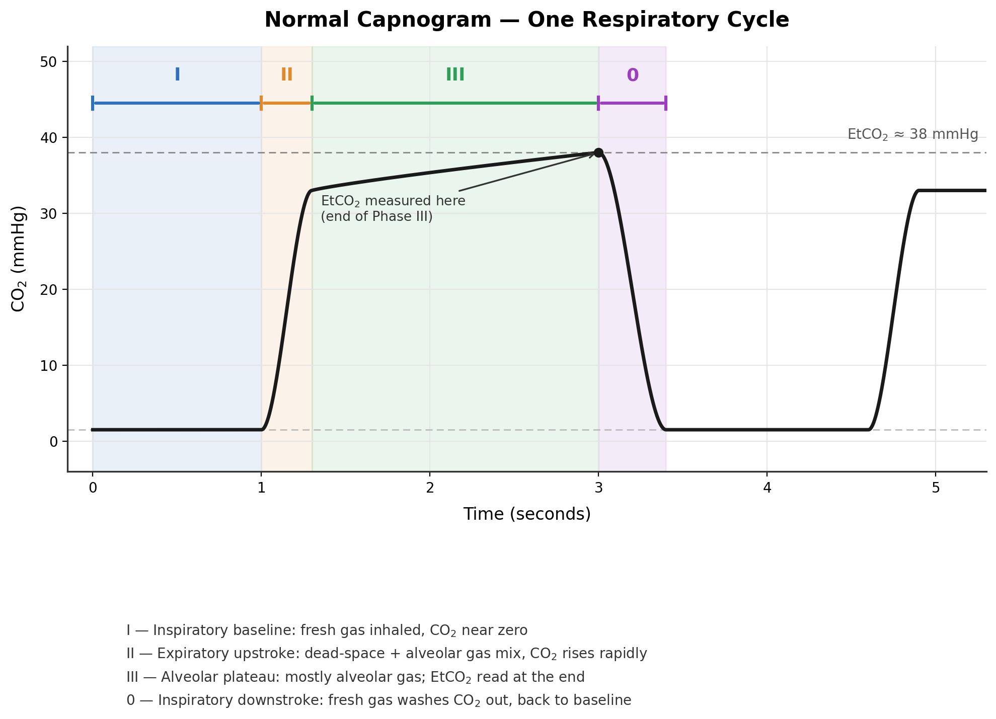
A normal capnogram has four recognizable phases:
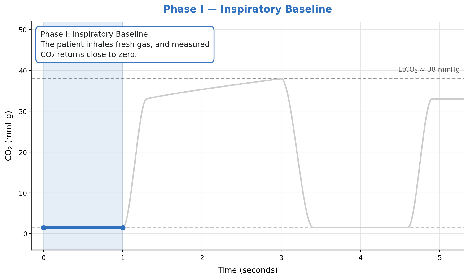
1. Phase I: Inspiratory baseline
The patient inhales fresh gas, and measured carbon dioxide should return close to zero.
2. Phase II: Expiratory upstroke
Exhalation begins. Gas from the conducting airways mixes with alveolar gas, producing a rapid rise in carbon dioxide.

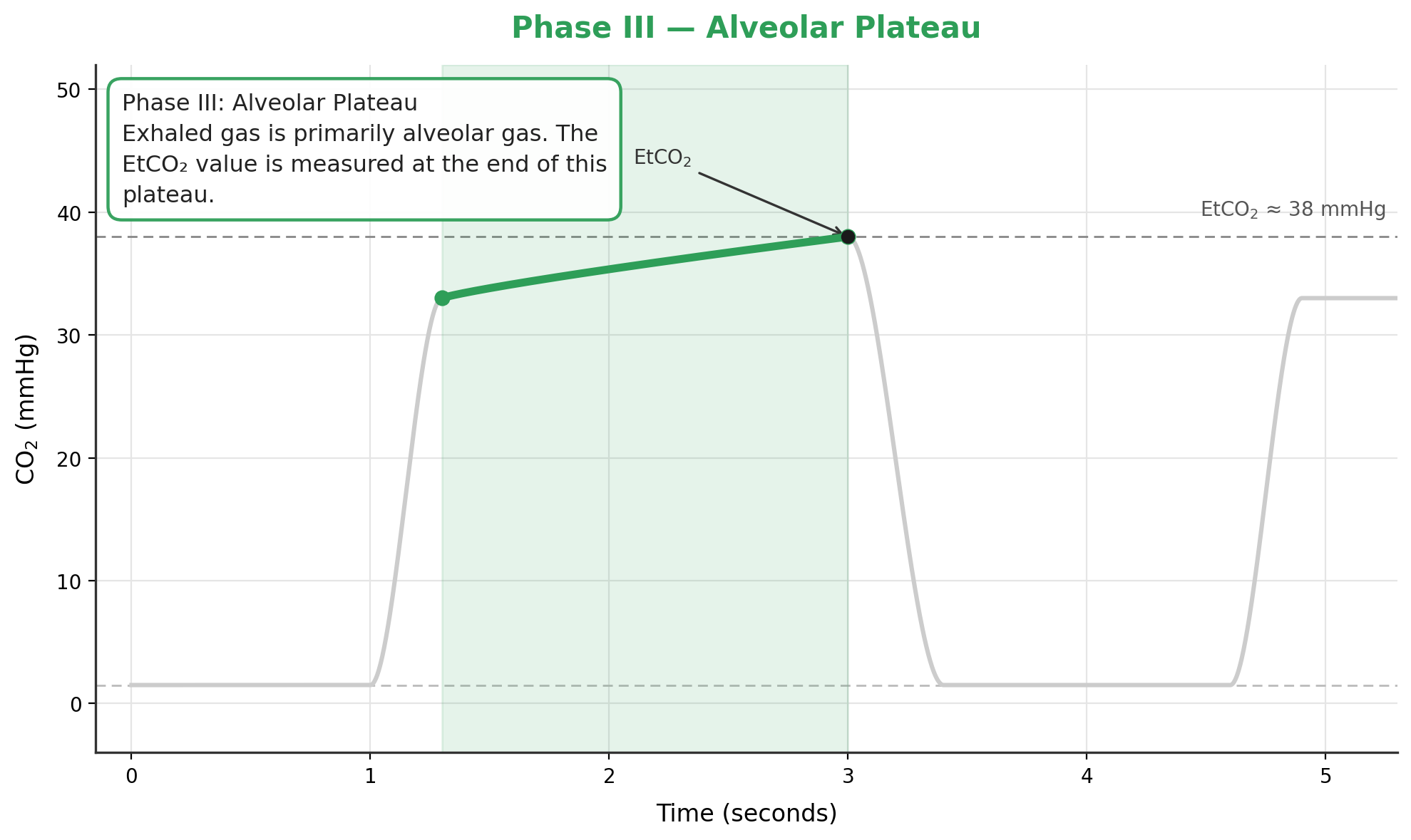
3. Phase III: Alveolar plateau
Exhaled gas is primarily alveolar gas. The EtCO₂ value is measured at the end of this plateau.
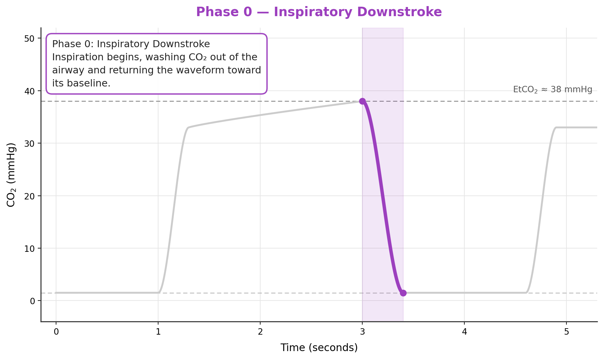
4. Phase 0: Inspiratory downstroke
Inspiration begins, washing carbon dioxide out of the airway and returning the waveform toward its baseline.
A stable capnogram generally has a near-zero baseline, a steep expiratory upstroke, a relatively level plateau, and consistent breath-to-breath timing. Waveform shape should always be interpreted alongside the numeric EtCO₂ trend and the patient’s clinical condition.
Mainstream and Sidestream Capnography
Capnography systems generally use either a sensor positioned directly at the airway or a system that draws a small gas sample through tubing to a sensor located within or connected to the monitor.
Mainstream capnography
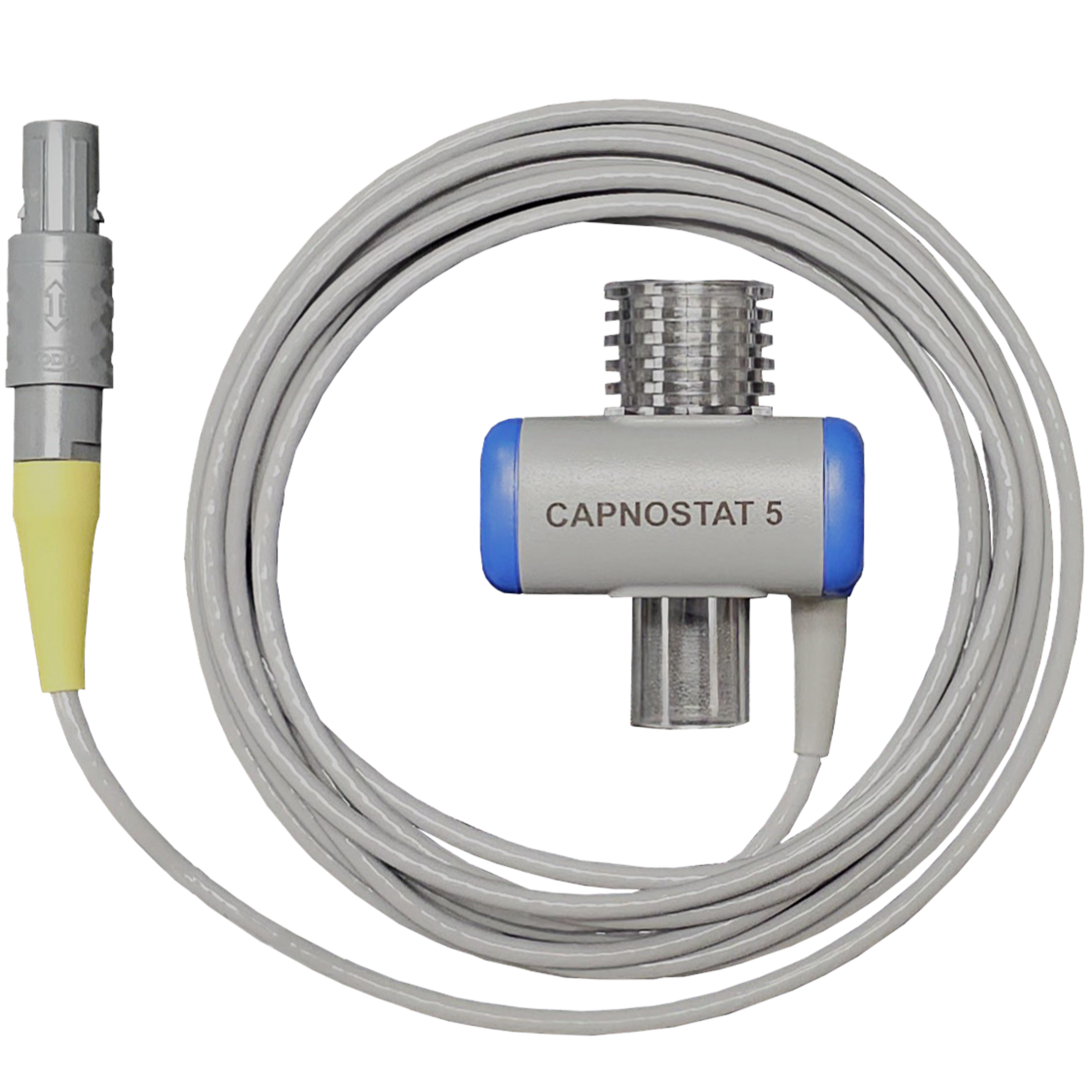
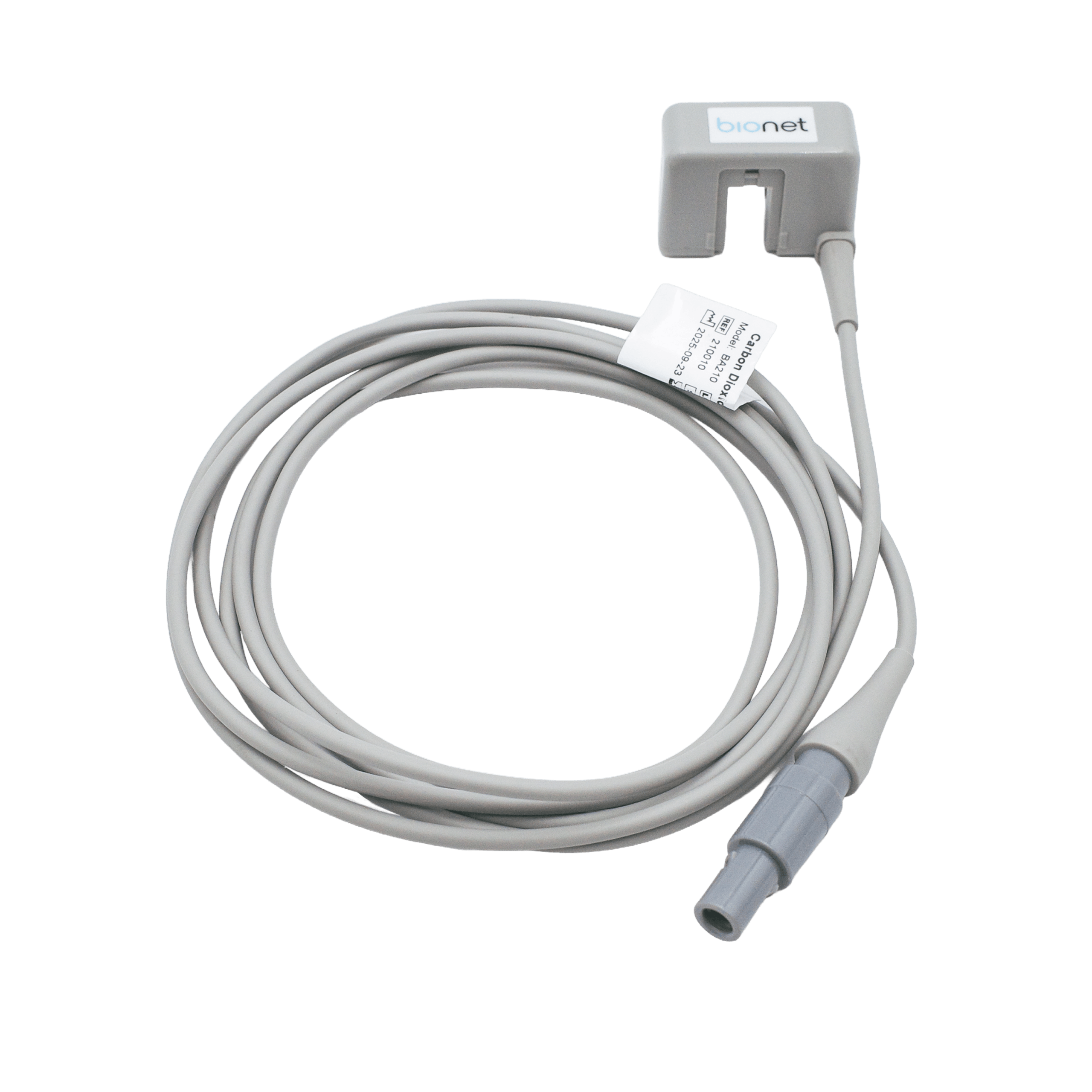
With mainstream capnography, the infrared sensor is positioned at the airway adapter. This provides rapid measurement and avoids the delay associated with transporting a sample through tubing.
Practical considerations include:
- The sensor and adapter add weight at the endotracheal tube.
- The adapter contributes mechanical dead space.
- The assembly should be supported so it does not pull on or displace the tube.
- Protect the sensor from fluid intrusion and excessive contamination, and handle and clean it according to the manufacturer’s instructions.
- The adapter may require zeroing according to the manufacturer’s instructions.
Bionet offers mainstream EtCO₂ monitoring for compatible Brio XVet configurations, allowing carbon dioxide to be measured directly at the patient’s airway.
Sidestream capnography
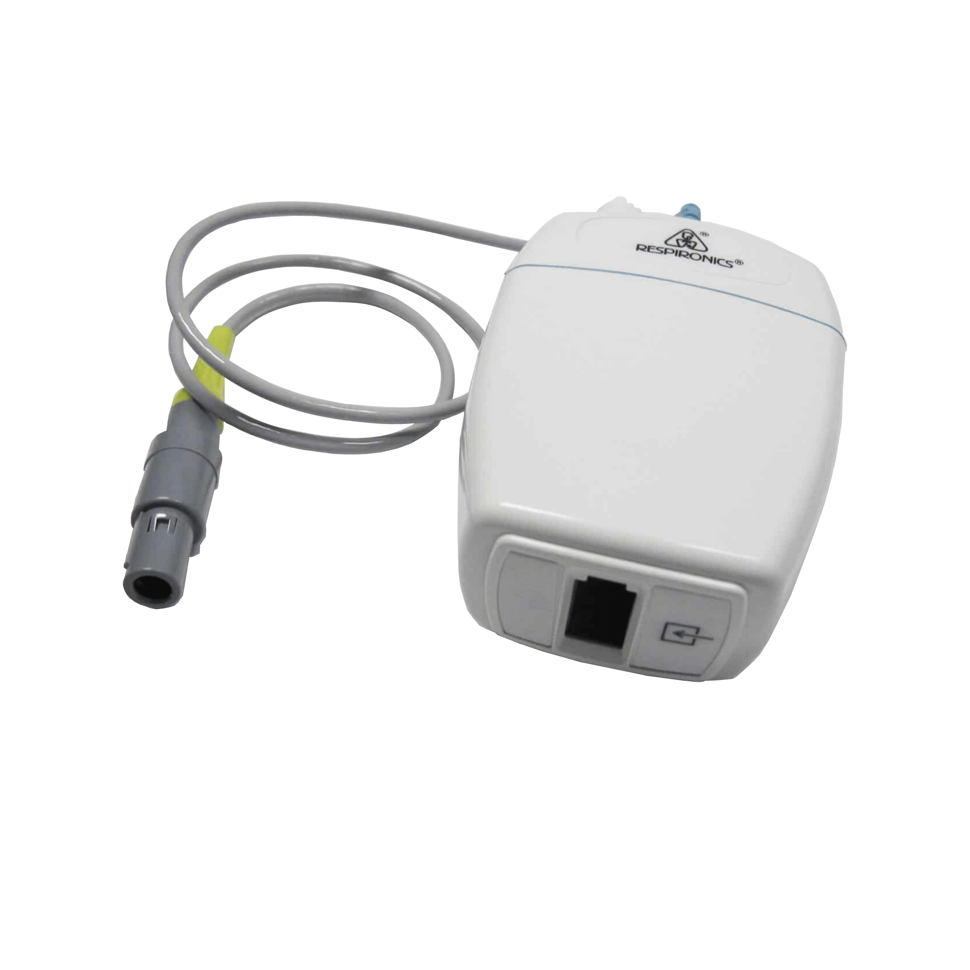

With sidestream capnography, a small volume of respiratory gas travels through a sampling line to a sensor located within or connected to the monitor. This configuration can reduce weight at the patient connection and may be convenient when space or patient positioning is a concern.
The clinical team should account for:
- A short measurement delay
- Sampling-line leaks, kinks, or disconnections
- Moisture accumulation
- Water-trap condition
- Correct line placement and secure connections
- In very small patients or patients with rapid respiratory rates and low tidal volumes, sampling flow, sampling-line volume, leaks, and response time may affect waveform quality and measurement accuracy. Use only patient interfaces and sampling components approved for the specific system.
Adapter Selection and Mechanical Dead Space
Mechanical dead space is the volume within equipment where inhaled and exhaled gas can mix without participating in gas exchange. It may include airway adapters, elbow connectors, Y-pieces, and sections of tubing beyond the patient’s anatomic dead space.
Mechanical dead space is especially important in:
- Cats
- Toy-breed and small dogs
- Pediatric patients
- Patients with respiratory compromise
Use a low-dead-space adapter appropriate for the patient and endotracheal tube. The adapter’s internal diameter should exceed the internal diameter of the endotracheal tube to avoid adding airway resistance. AAHA recommends limiting total equipment dead space to approximately 2–3 mL/kg whenever possible.[3]
Always confirm sizing limits in the manufacturer’s instructions. A smaller patient does not simply require a smaller version of the same setup: every added component should be considered as part of the patient’s total breathing workload.
Capnography Setup and Monitoring Checklist
Before anesthesia
- Confirm the patient’s history, physical examination, and anesthetic plan.
- Select the appropriate endotracheal tube, circuit, adapter, and sampling configuration.
- Inspect the capnograph monitor, sensor, cable, adapter, sampling line, and water trap.
- Check for cracks, kinks, moisture, occlusions, and loose fittings.
- Turn on the monitor and capnography module.
- Zero or calibrate the system only as directed by the manufacturer and away from the patient’s carbon dioxide source.
- Perform the anesthesia-machine leak and pressure checks.
- Confirm oxygen supply, vaporizer function, absorbent condition, valves, pop-off valve, and ventilator readiness.
- Set appropriate alarms and verify that alarm volume is audible.
- Prepare backup airway and ventilation equipment.
During anesthesia
- Confirm a persistent waveform after intubation.
- Observe the patient, reservoir bag or ventilator bellows, chest movement, airway pressure, and breathing pattern.
- Trend EtCO₂ rather than reacting to one isolated number.
- Review waveform shape with every meaningful change in EtCO₂.
- Record EtCO₂, heart rate, blood pressure, oxygen saturation, and other required parameters according to hospital protocol.
- Keep the sampling line free of moisture and tension.
- Escalate abnormal findings to the veterinarian promptly.
After anesthesia
- Continue monitoring ventilation and oxygenation during recovery.
- Assess airway patency, respiratory effort, chest excursion, and mucous membrane color.
- Continue capnography when clinically indicated and equipment is compatible with the recovery setup.
- Document abnormal values, waveform changes, interventions, and patient response.
- Maintain observation until the patient is physiologically stable according to the attending veterinarian’s criteria.
Common EtCO₂ and Waveform Abnormalities
These patterns are clinical clues, not diagnoses. Every abnormality requires correlation with the patient and the complete monitoring picture.
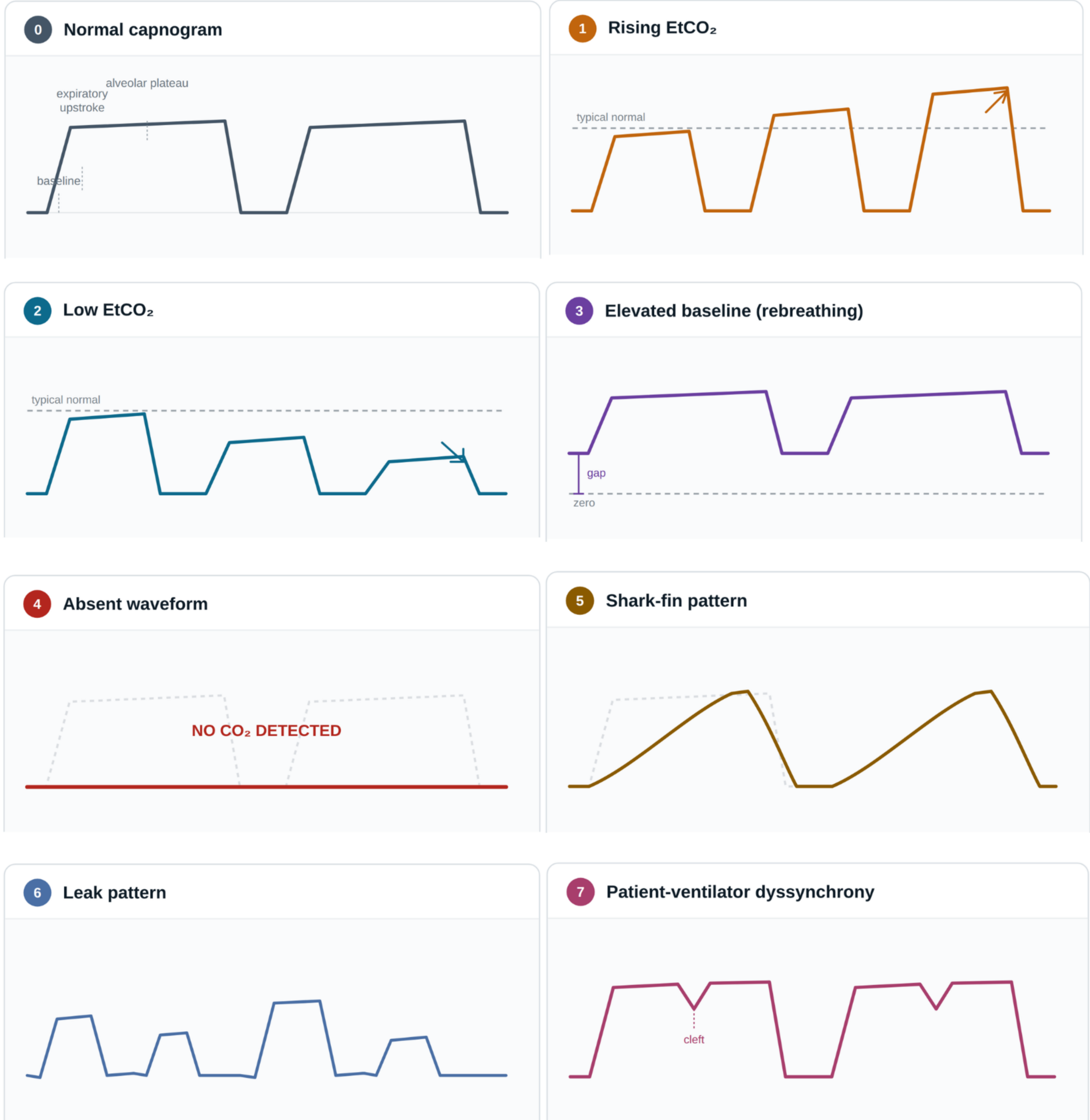
- Rising EtCO₂ with a recognizable waveform: May indicate hypoventilation, excessive anesthetic depth, inadequate ventilator support, increased dead space, or increased carbon dioxide production.
- Low EtCO₂: May reflect hyperventilation, reduced pulmonary perfusion, low cardiac output, hypotension, leaks, or poor sampling.
- Elevated inspired CO₂ or a baseline above zero: Suggests rebreathing. Possible causes depend on the breathing circuit. In a rebreathing circuit, consider exhausted CO₂ absorbent or malfunctioning one-way valves. In a nonrebreathing circuit, consider inadequate fresh-gas flow. Also inspect for excessive equipment dead space or other circuit problems.[3]
- Absent waveform: Treat as an emergency until proven otherwise. Consider apnea, esophageal intubation, accidental extubation, circuit disconnection, tube occlusion, sampling-line obstruction, or monitor failure.
- Shark-fin pattern: A slanted upstroke and poorly defined plateau may suggest partial airway obstruction, a kinked or obstructed tube, bronchospasm, or increased airway resistance.
- Leak pattern: An irregular or shortened waveform may result from an endotracheal-tube leak, circuit leak, loose adapter, or disconnected component.
- Patient-ventilator dyssynchrony: Small notches or clefts in the waveform may indicate spontaneous respiratory effort against mechanical ventilation. Assess anesthetic depth, pain, temperature, carbon dioxide, oxygenation, and thoracic restriction.
A Systematic Troubleshooting Approach
When the waveform or EtCO₂ changes unexpectedly, use a consistent sequence:
- Assess the patient first. Check airway, breathing, circulation, anesthetic depth, chest movement, pulse, heart rate, blood pressure, and oxygen saturation.
- Check the endotracheal tube. Confirm position, patency, cuff seal, kinks, mucus, and connection.
- Check the breathing circuit. Inspect limbs, connectors, valves, pop-off valve, ventilator, and circuit integrity.
- Check the sampling pathway. Look for leaks, cracks, occlusion, moisture, and loose connections.
- Check the water trap. Replace water trap once it is full
- Check the monitor and sensor. Confirm power, module connection, zeroing requirements, alarm settings, and manufacturer guidance.
- Reassess after intervention. Confirm that the waveform and patient parameters respond appropriately.
Integrating Capnography with Complete Anesthesia Monitoring
Capnography should not be used alone. Combine it with:
- SpO₂ for oxygenation
- ECG for heart rate and rhythm
- Blood pressure for perfusion assessment
- Respiratory rate and chest excursion
- Airway pressure or ventilator data
- Temperature
- Anesthetic depth
- Inspired and expired anesthetic agent concentrations
- Arterial or venous blood gas analysis when indicated
Blood gas analysis may be particularly valuable when there is significant pulmonary disease, ventilation-perfusion mismatch, abnormal ventilation before anesthesia, thoracic disease, or a persistent discrepancy between EtCO₂ and the patient’s clinical condition.
Bionet Solutions for Veterinary Capnography
Bionet’s veterinary monitoring platform supports flexible capnography workflows. The Brio XVet multiparameter monitor lineup includes the Brio X3Vet, Brio X5Vet, and Brio X7Vet, with compatible EtCO₂ configurations for mainstream or sidestream capnography monitoring , or Dual Gas for capnography and anesthetic agent monitoring for even more valuable anesthesia values.
Bionet’s Mainstream and Sidestream EtCO₂ options measure carbon dioxide and provides continuous EtCO₂, inspired CO₂, respiratory rate, and waveform monitoring. The Dual Gas module adds EtCO₂ and provides portable EtCO₂, inspired CO₂, respiratory rate, anesthetic agent concentration, and MAC values for selected anesthetic agent measurements to compatible Brio XVet systems.
Brio XVet monitors include a four-year monitor warranty, touchscreen operation, data-management capabilities, free remote training, and access to Bionet America’s U.S.-based technical-support team. Mainstream and Sidestream Capnography modules carry a 2 year warranty.
Quick-Reference Summary
- Confirm the correct patient interface, adapter, and monitoring configuration for the patient’s size and airway setup.
- Minimize equipment dead space and avoid unnecessary connectors.[3]
- Confirm a persistent waveform after intubation and trend both EtCO₂ and waveform shape.
- If values change unexpectedly, assess the patient first, then the airway, circuit, sampling pathway, absorbent or flow source, and monitor.
- Use capnography alongside SpO₂, ECG, blood pressure, temperature, anesthetic depth, and blood gas analysis when indicated.
Frequently Asked Questions
What is a typical EtCO₂ range during anesthesia?
Reference ranges vary by patient, protocol, ventilation method, and clinical condition. AAHA guidance describes approximately 40–50 mmHg as common in appropriately anesthetized dogs and cats, with values up to about 55 mmHg potentially tolerated in selected patients.[4] Trends and clinical context are essential.
Why might PaCO₂ and EtCO₂ differ?
Is capnography a replacement for pulse oximetry?
No. Capnography evaluates ventilation and carbon dioxide elimination, while pulse oximetry evaluates oxygen saturation. Both provide different and complementary information.
To discuss the right EtCO₂ configuration for your practice, contact Bionet for more information.
Built for Modern Veterinary Machine
Smarter Solutions • Smarter Care • Better Outcomes
References
- A Guide to Capnography in Small Animal Anesthesia : Today’s Veterinary Nurse
- 2025 ACVAA Small Animal Anesthesia and Sedation Monitoring Guidelines
- 2020 AAHA Anesthesia and Monitoring Guidelines for Dogs and Cats
- AAHA Guidance: Hypoventilation
- AAHA Guidance: Equipment Preparation
- Bionet Brio X Series Veterinary Multiparameter Monitors
- Bionet Veterinary Capnography Solutions
- Bionet GA3VET EtCO₂/Anesthetic Gas Analyzer
- Bionet Dual Gas Module